Could Brain Scans Diagnose this Mysterious Disease?
Could Brain Scans Diagnose this Mysterious Disease?
March 27, 2014
During the era of flappers and prohibition speakeasies, physicians in the United States discovered a mysterious neurodegenerative disorder while conducting post-mortem exams on boxers. Today, the disease is known as Chronic Traumatic Encephalopathy (CTE), a progressive and deteriorating disease that develops in the brains of athletes with a history of repetitive brain trauma.
Changes within the brain due to CTE can begin years or even decades after a brain trauma event, making it difficult to track. The degeneration of the brain tissue is associated with depression, memory loss and, ultimately, dementia.
Late last May, Cullen Finnerty, a 30-year-old former college football quarterback, went fishing by himself in a patch of Michigan woods. He had complained of headaches and difficulty sleeping before he left. After he was missing for several days, he was found dead. An autopsy later revealed that Finnerty suffered from CTE — though it’s not clear how it was connected to his death.
While some 90 or so years have passed since CTE was first found, medical science has progressed little in its understanding of the disease. Physicians are still able to conclusively diagnose a case only after a suspected sufferer has died.
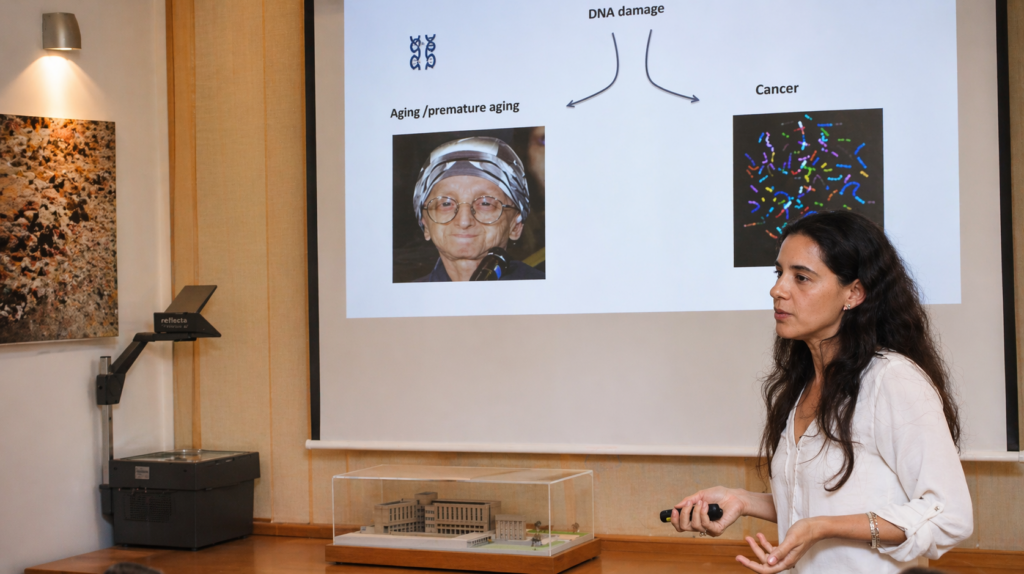
“When you’re diagnosed it’s too late,” says Prof. Alon Friedman, chair of BGU’s Zlotowski Center for Neuroscience and head of BGU’s Laboratory for Experimental Neurosurgery, a collaboration with Soroka University Medical Center.
This makes it near impossible to estimate who and how many people are at risk of CTE. Not only is it problematic to diagnose CTE, we also know remarkably little about its pathology.
Prof. Friedman is trying to solve this problem by scanning the human brain using magnetic resonance imaging (MRI) technology. He and his team of researchers at BGU’s Brain Imaging Research Center hope to find a way to diagnose the disease before significant loss of brain tissue occurs.
They aim to detect leakage in the blood-brain barrier (BBB), which protects the brain’s grey matter from toxins in the blood system that are harmful to the brain.
Prof. Friedman has modified a technique called dynamic contrast enhanced MRI. The technique requires researchers to conduct an initial MRI scan, which they then compare to a series of scans after they’ve intravenously injected a contrast agent into the bloodstream.
If the BBB is compromised, the contrast agent will trickle into the brain and show up on screen, while a healthy brain shows no change in magnetic characteristics.
The process allows doctors to quantify BBB damage by comparing the scans with and without the contrast agent, which Prof. Friedman hopes to use as a diagnostic tool. “We can measure and give this change a number — reflecting how much of it was leaking.”
Prof. Friedman says it would be progress to see his methods routinely implemented, at least for patients who are highly susceptible to leakage. That, he says, could finally bring the promise of earlier diagnosis into the realm of possibility.
Read more on the Dell-sponsored page of The New York Times website >>
Stories Like This